 |
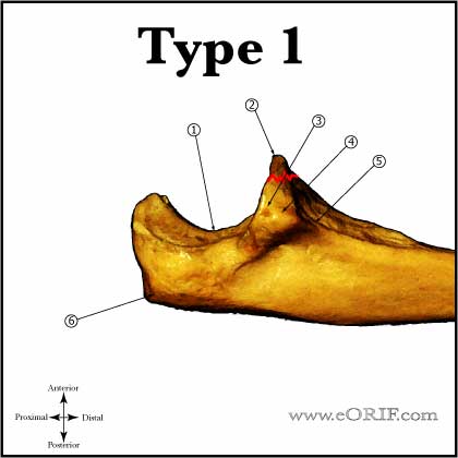
Type I Coronoid Fracture, stable (avulsion of the tip of the coronoid process): usually related to posterolateral rotatory elbow subluxation. Treatment = early protected ROM. Type I, unstable or associated radial head fracture. Treatment = ORIF if fragment is large enough for fixation with a screw or k-wire. Type I coronoid fractures result in only small changes in elbow kinematics are not corrected with suture repair. Any collateral ligament injury must be repaired as well. Medial view of Proximal Ulna
|
 |
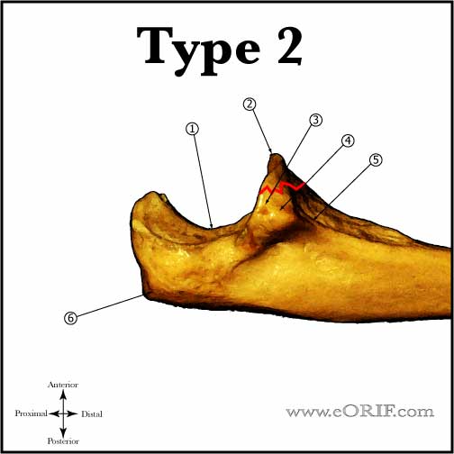
Type II Coronoid Fracture, stable( <50% of coronoid): Treatment = early protected ROM. Type II, unstable or associated radial head fracture. Treatment = ORIF; if fragment is large enough for fixation with a screw or k-wire it should be fixed. Any collateral ligament injury must be repaired as well. Medial view of Proximal Ulna
|
 |
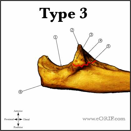
Type III Coronoid Fracture (basal coronoid fracture): Treatment = ORIF usually via a posteromedial aproach. Often associated with olecranon fracture/dislocations. Associated injuries should be anatomically repaired as well. Always consider hinged external fixation for severe injuries where joint stability is a concern post-operatively. Medial view of Proximal Ulna
|
 |
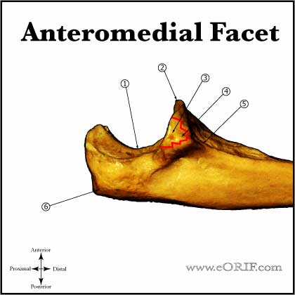
Anteromedial Facet Coronoid Fracture, stable: Occur with varus posteromedial rotation during axial loading. Associated with LCL rupture and are usually unstable. Treatment = ensure joint is stable with stress radiographs, consider EUA. Early protected ROM if joint is confirmed to be stable. Anteromedial facet fracture, unstable: Treatment = ORIF with concomitant LCL/radial head repair usually via a utilitarian posterior exposure with posteromedial coronoid exposure. Always consider hinged external fixation for severe injuries where joint stability is a concern post-operatively. Medial view of Proximal Ulna
|
|
Coronoid Review References |
