|
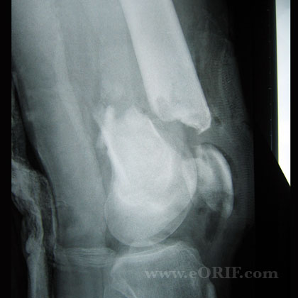
Synonyms: Supracondylar femur fracture, distal femur fracture
Distal Femur Fracture ICD-10
A- initial encounter for closed fracture
B- initial encounter for open fracture type I or II
C- initial encounter for open fracture type IIIA, IIIB, or IIIC
D- subsequent encounter for closed fracture with routine healing
E- subsequent encounter for open fracture type I or II with routine healing
F- subsequent encounter for open fracture type IIIA, IIIB, or IIIC with routine healing
G- subsequent encounter for closed fracture with delayed healing
H- subsequent encounter for open fracture type I or II with delayed healing
J- subsequent encounter for open fracture type IIIA, IIIB, or IIIC with delayed healing
K- subsequent encounter for closed fracture with nonunion
M- subsequent encounter for open fracture type I or II with nonunion
N- subsequent encounter for open fracture type IIIA, IIIB, or IIIC with nonunion
P- subsequent encounter for closed fracture with malunion
Q- subsequent encounter for open fracture type I or II with malunion
R- subsequent encounter for open fracture type IIIA, IIIB, or IIIC with malunion
S- sequela
Distal Femur Fracture ICD-9
- 821.23(closed)
- 821.33(open)
Distal Femur Fracture Etiology / Epidemiology / Natural History
- High-energy (MVA, Falls from height) in young patients, fragility fracture in elderly patients, periprosthetic fracture.
Distal Femur Fracture Anatomy
- Hoffa Fragment = coronal (frontal) plane fragment associated with comminution in the intercondylar notch. Present in @1/3 of Type C fractures.
Distal Femur Fracture Clinical Evaluation
- ATLS resuscitation. These can be high enegery injuries, assessment should begin with the A,B,C's.
- Obvious deformity of knee/thigh often with limb shortening
- Document neurovascular exam before and after any treatment.
Distal Femur Fracture Xray
- A/P and lateral views of the knee.
- CT: Ct scan is nearly always indicated for pre-operative planning as there is a high association with coronal plane fractures which are difficult to see on plane films. (Nork SE, JBJS Am 2005;87:564-569)
Distal Femur Fracture Classification/Treatment
- AO Classification(link is external)
- Type A=extraarticular
-Treatment = retrograde IM nail or ORIF usually via minimal lateral approach
- Type B=unicondylar fractures
-Treatment = percutaneous lag screw fixation +/- plating
- Type C=intrarticular fractures
-Treatment = ORIF via modifed lateral parapatellar arthrotomy (Starr AJ, JOT 1999;13:138)(link is external). Generally articular reduction and submuscular locked plate positioning via a small incision with spanning of metaphyseal comminution.
- Periprosthetic distal femur fracture (Periprosthetic supracondyle femur fracture)
-Treatment = Lateral locked plating with indirect reduction (Ricci WM, JOT 2006;20:190)(link is external). Poor bone stock, loose prosthesis, or PS-TKA consider distal femoral replacement. (Kassab, M J Arthroplasty 2004;19:361-368)
- Consider primary TKA with tumor prosthesis for elderly osteoporotic patients with severe comminution. The linea aspera is the key to rotational alignment during surgery. Drape out normal leg and ensure femoral head can be visualized with c-ram.
Distal Femur Fracture Associated Injuries / Differential Diagnosis
- Multiply injured patient
- Meniscal tear
- Knee ligmanetous injury: ACL / MCL / LCL / PCL / PLC.
Distal Femur Fracture Complications
- Nonuion
- Malunion
- Infection
- Failure of fixation
- Painful hardware
- Compartment Syndrome
- CRPS
- DVT / PE
- Heterotopic Ossification
- Extension contracture: uncommon, usually caused by periarticular fibrosis and shortening of the rectus femorus. The Judet quadricepsplasty, based on muscle disinsertion and sliding, is the treatment of choice (Ebraheim NA, J Orthop Trauma 1993;7:327-330).
Distal Femur Fracture Follow up care
Distal Femur Fracture References
|


