 |
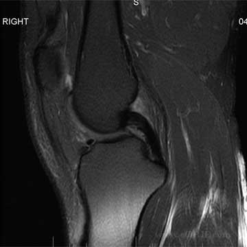
PCL MRI - Normal
- 96-100% accurate in determining all posterior cruciate ligament (PCL) injuries. (Fischer JBJS 73A:2-10 1991).
- PCL appears as thick dark band extending from the medial femoral condyle to the posterior tibia best seen on oblique sagittal images.
- PCL disruption = discontinuity, abnormal course, or fluid signal traversing the ligament on T2 images.
|
 |
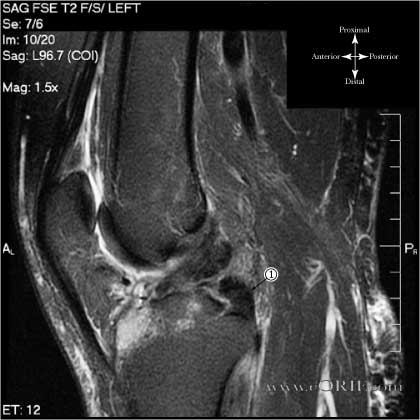
PCL Tear
- Sagittal FSE T2 MRI of 28y/o male with PCL and ACL tears.
- Image through the intercondylar notch demonstrating torn PCL shown.
- Stump of PCL demonstrating discontinuity with origin on medial femoral condyle and lax, abnormal contour.
|
 |
Bucket Handle Meniscal Tear
- Coronal T1 MRI of bucket handle lateral meniscal tear
- Medial Meniscus
- Bucket handle lateral meniscal tear displaced into the intercondylar notch
- Absent lateral meniscus in normal position
|
 |
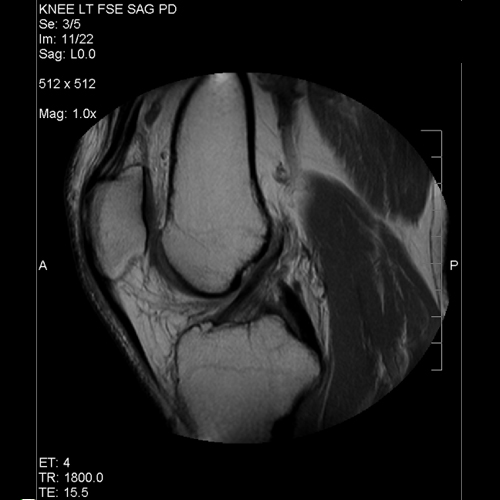
Proton Density MRI of Normal ACL
- Quadriceps tendon
- Patella
- Patellar ligmanet
- Distal femur (notch area)
- ACL
- Tibial Plateau
- PCL
Sagittal FSE PD image
|
 |
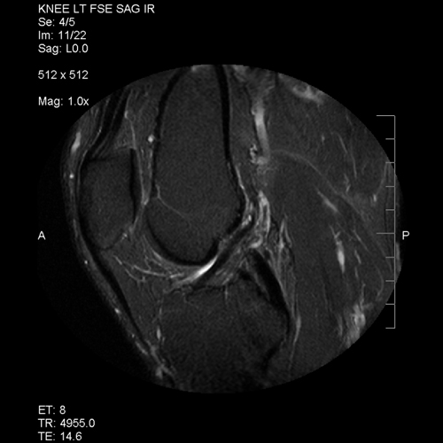
IR MRI of normal ACL |
| |
|


