|
|
synonyms: medial epicondyle fracture
Medial Epicondyle Fx ICD-10
A- initial encounter for closed fracture
B- initial encounter for open fracture
D- subsequent encounter for fracture with routine healing
G- subsequent encounter for fracture with delayed healing
K- subsequent encounter for fracture with nonunion
P- subsequent encounter for fracture with malunion
S- sequela
Medial Epicondyle Fx ICD-9
- 812.43 (closed medial condyle fracture)
- 812.53(open)
Medial Epicondyle Fx Etiology / Epidemiology / Natural History
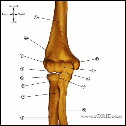
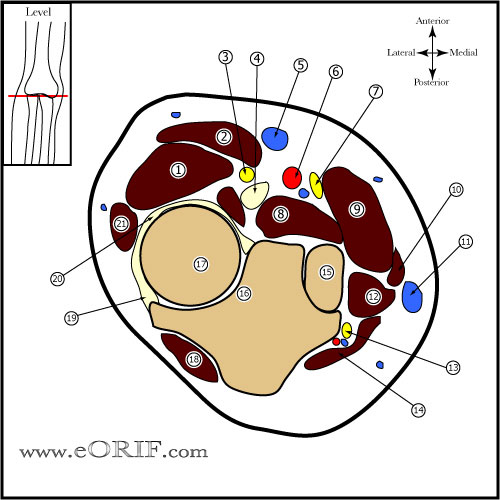
Medial Epicondyle Fx Anatomy
- Primary constraints=ulnohumeral articulation(coronoid), medial collateral ligament(MCL), lateral collateral ligament(LCL) (King GJ, JSES 2;165:1993).
- Secondary constraints=radial head, common flexor and extensor origins, capsule. (Morrey BF, CORR 1991;265:187).
- Dynamic constaints=mucles which cross the elbow, mainly the triceps, anconeus and brachialis.
- Anterior band of medial collateral ligament is the primary constraint to valgus instability.
- LCL(ulnar part) is primary constraint to posterolateral rotatory instability
- Pathoanatomy of dislocation is a circle of bone/soft tissue disruption starting laterally and progressing medially. Stage I=ulnar band of LCL, Stage II=ant/post capsule, Stage III=MCL disrupted, anterior band of MCL is the last to disrupt.
- Most dislocations have disruptions of both the MCL and LCL.
- (McKee MD, JSES 2003;12:391).
- See also Elbow Anatomy.
Medial Epicondyle Fx Clinical Evaluation
- Generally present with obvious deformity, pain and swelling.
- Document NV exam.
- Lateral pivot shift test=for posterolateral rotatory instability- pt supine, arm overhead. Supination-valgus moment applied during flexion, elbow subluxates usually at @40degrees, additional flexion causes reduction/clunk. Should create apprehension.
- Valgus and varus stress, both in extension and 30 degrees flexion.
- Valgus stress testing performed in full pronation to eliminated posterolateral rotatory instability.
- Document wrist evaluation.
Medial Epicondyle Fx Xray / Diagnositc Tests
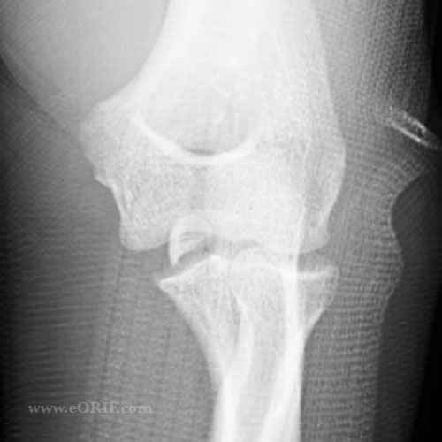
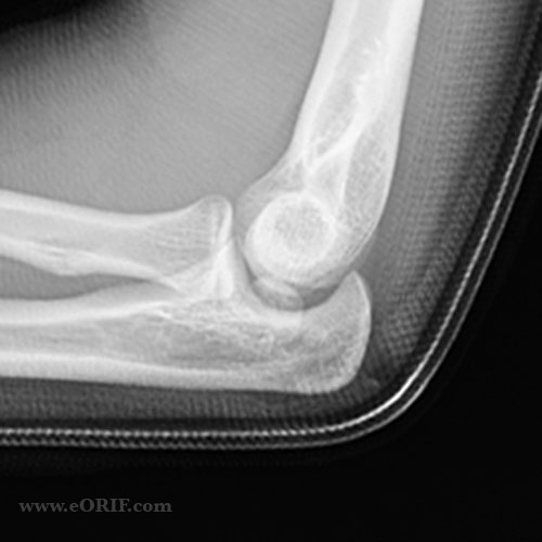
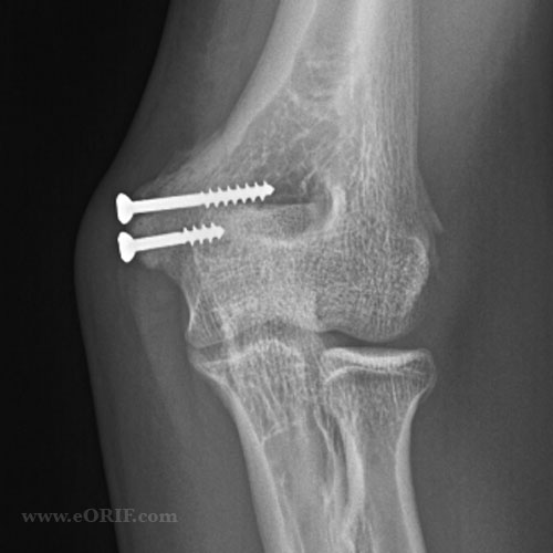
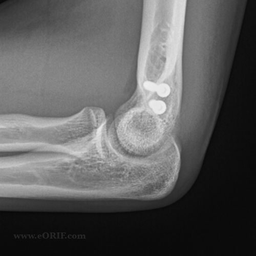
- A/P and lateral elbow films demostrates dislocation and associated fractures.
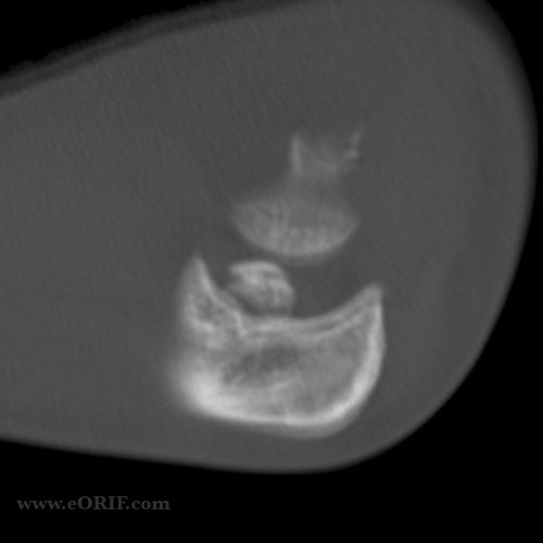
- Consider CT scan if fracture fragment is not clearly seen on xray.
Medial Epicondyle Fx Classification / Treatment
- Humeral Condyle CPT Coding(link is external)
- 24566 (percutaneous fixation of humeral epicondylar fracture, medial or lateral , with manipulation)
- 24575 (open treatment of humeral epicondylar fracture, medial or lateral, with or without internal or external fixation)
- 24582 (percutaneous fixation of humeral condylar fracture, medial or lateral , with manipulation)
- 24579 (open treatment of humeral condylar fracture, medial or lateral, with or without internal or external fixation)
- 24586 (open treatment of periarticular fracture and/or dislocation of the elbow; fracture distal humerus and proximal ulna and or proximal radius)
Medial Epicondyle Fx Associated Injuries / Differential Diagnosis
Medial Epicondyle Fx Complications
Medial Epicondyle Fx Follow-up Care
- Bulkly dressing with posterior splint post-operatively
- 7-10 day post-operative: Splint removed, ROM in a hinged elbow brace is started with ROM determined by security of fixation achieved at surgery.
- See also Elbow Outcome Measures.
Medial Epicondyle Fx Review References
|


