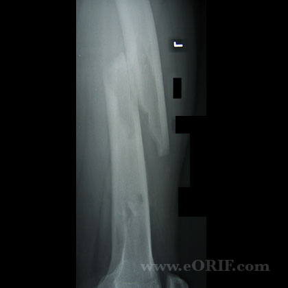
synonyms:femoral shaft fracture external fixation, ex fix
Femoral Shaft External Fixation CPT
Femoral Shaft External Fixation Indications
- Multi-trauma, Unstable patient (hypotensive, elevated lactate, base deficit, pulmonary injury, brain injury
- Open Grade III B & Cfemoral shaft fracture
- Bilateral femoral shaft fracture
Femoral Shaft External Fixation Contraindications
Femoral Shaft External Fixation Alternatives
- Traction: time to union can be 12 weeks.
Femoral Shaft External Fixation Pre-op Planning
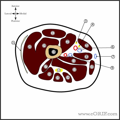
- Risks=femoral vessels, femoral nerve, sciatic nerve and its divisions, hip&knee joints
- Deep femoral vessels and saphenous nerve are most vulnerable as they pass through Hunter’s canal which can extend from the mid-aspect of the femur to the distal 1/6th.
- Knee joint(suprapatellar pouch) extends anterior @6.3cm above the proximal pole of the patella
Femoral Shaft External Fixation Technique
- Sign operative site.
- Pre-operative antibiotics.
- General endotracheal anesthesia
- Supine position on radiolucent OR table. All bony prominences well padded.
- Examination under anesthesia of the knee.
- Ensure adequate A/P and lateral c-arm views can easily be obtained
- Prep and drape in standard sterile fashion.
- Safe corridor is mainly anterolateral, between a line medial aspect of the medial femoral condyle to the ASIS and a line from the mid-aspect of the intertroch area to the most lateral aspect of the lateral femoral condyle.
- Avoid pins in rectus femoris to avoid soft-tissue adhesions and knee stiffness.
- Generally use 5- 6mm half pins for adults, 4-5mm for children. Pin diameter should not exceed 1/3 the diameter of the bone at the insertion site.
- Place two pins proximal to fracture and two pins distal.
- Construct fixator per manufactor technique.
- Applied traction to reduce fracture and tighten all points on fixator
- Dress pins.
Femoral Shaft External Fixation Complications
- Infection-0.9%
- Nonuion=0.9%
- Malunion(rotation,shortening,angulation) rotational difference > 15 degrees from unaffected side is a true malrotation deformity. Hip and knee symptoms and functional disability occur when rotation is >20 degrees. Rotational differences <10 degrees seldom produce symptomatic disability.
- Peroneal nerve palsy
- Fat-embolism sydrome
- ARDS
- DVT / Pulmonary Embolism
- Pudendal Nerve palsy: compression of the pudendal nerve from traction against the perineal post on the fracture table in the supine position can lead to perineal numbness following intramedullary nailing. Attention to detail in placement of the patient and adequate padding can minimize this risk. (Brumback RJ,.JBJS 1992;74A:1450-1455)
- DIC
- Compartment Syndrome
- Heterotopic Ossification
- Knee pain (retrograde nail)
- Thigh pain (antegrade nail)
- Abductor lurch (antegrade nail)
Femoral Shaft External Fixation Follow-up care
- Post-op:
- 7-10 Days:
- External fixation generally converted to IM nail within 2weeks of application to decrease deep infection risk (Nowotarski PJ, JBJS 2000;82A:781),(Harwood PJ, JOT 2006;20:181).
Femoral Shaft External Fixation Outcomes
Femoral Shaft External Fixation Review References
- Rockwood and Greens
- Broos PL, Miserez MJ, Rommens PM: The monofixator in the primary stabilization of femoral shaft fractures in multiply-injured patients. Injury 1992;23:525-528.
- Kishan S, Techniques in Orthopaedics, 17(2):239-244, 2002) (Scalea, J Trauma 48:613;2000
- °


