 |
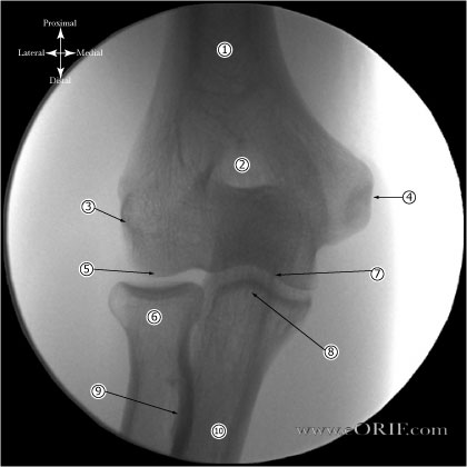
A/P Elbow Xray - Forearm in suppination
- Humeral Shaft
- Olecranon fossa
- Lateral epicondyle
- Medial epicondyle
- Capitellum
- Radial head
- Trochlea
- Conoid tubercle
- Radial tuberosity
- Ulnar Shaft
|
 |
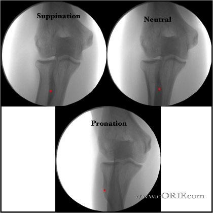
A/P Elbow through a ROM from full suppination to full pronation.
Red dot indicates position of radial tuberosity which moves from:
- Anterior position in suppination
- Posteromedial in neutral rotation
- Lateral in pronation
|
 |
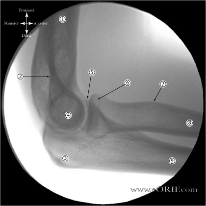
Lateral Elbow Xray- Forearm in suppination
- Humeral Shaft
- Olecranon fossa
- Olecranon
- Medial and lateral epicondyle overlapping
- Conoid tubercle
- Radial head
- Radial uberosity
- Radial shaft
- Ulnar shaft
|
 |
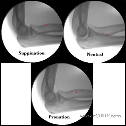
Lateral Elbow through a ROMfrom full suppination to full pronation.
Red dot indicates position of radial tuberosity which moves from:
- Anterior position in suppination
- Posteromedial in neutral rotation
- Lateral in pronation
|
| |
Radial Head-Capitellum view
- Demostrates: Unobstructed view of radial head, capitellum, coronoid process, humeroradial/humeroulnar articulations • anteroinferior glenoid rim.
- Helpful for:
Postion: patient seated with forarm resting on the xray table on its ulnar side; elbow flexed 90°; thumb pointing up.
Beam: aimed at 45° angle from the forearm towards radial head
|
| |
Oblique - Olecranon osteophytes
An oblique axial view with the elbow in 110° of flexion best demonstrates posteromedial olecranon osteophytes.
Wilson FD, AJSM 1983;11: 83–88
|