|
|
synonyms:SLAC wrist, Scapho-Lunate Advanced Collapse, scapholunate advanced collapse
SLAC ICD-10
SLAC ICD-9
- 715.13 Osteoarthrosis, localized, primary, idipathic, forearm
SLAC Etiology / Epidemiology / Natural History
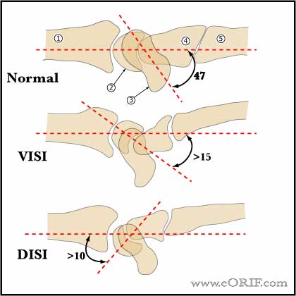
- Primary disorder in the SLAC wrist is that of scapholunate dissociation secondary to scapholunate interosseous ligament rupture . Scapholunate dissociation leads to unopposed volar flexion of the scaphoid and the dorsal intercalated segmental instability (DISI) pattern
- Scapholunate dissociation is generally from trauma, but may occur from calcium pyrophosphate deposition.
- Most common cause of wrist arthritis. 57% of degenerative wrist arthritis (Watson HR, Ballet FL. Journal of Hand Surgery 1984;9A, No. 3 May 1984).
SLAC Anatomy
SLAC Clinical Evaluation
- Variable degrees of wrist pain, swelling and decreased ROM. Advanced disease is associated with night pain.
- May have remote history of wrist trauma.
SLAC Xray / Diagnositc Tests
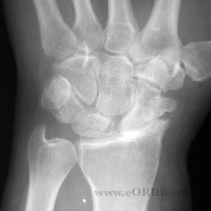
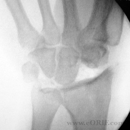
- Degenerative changes progress from the radial styloid and scaphoid along the scaphoradial joint.
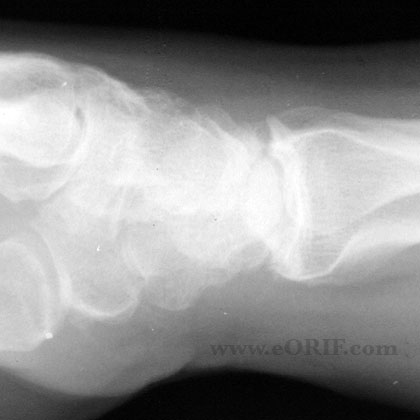
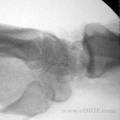
- Lateralradiographs may show the scapholunate angle to be increased beyond 60°, which is felt to be the upper limit of normal. Normal scapholunate angle=47 range=30-60 degrees.
- PAradiograph, the scaphoid appears foreshortened, has a “cortical ring” sign(volar flexed scaphoid distal pole seen in cross section) and there is a scapholunate gap of greater than 3 mm
- PA clenched fistview in ulnar deviation accentuates widenings at the scapholunate interval
- Carpal height Index= distance between the base of the third metacarpal and the articular surface of the radius divided by the length of the third metacarpal on a neutral P/A xrays. Normal = 0.54 +/- 0.03. Best evaluated by comparing Carpal height index to that of the normal side. (Mann Fa, Radiology 1992;184:15). Can also compare carpal height index using the height of the capitate. Normal using capitate = 1.57 +/- 0.05.
SLAC Classification / Treatment
SLAC Associated Injuries / Differential Diagnosis
- Preiser's Disease
- Scaphoid fracture
- Scaphoid nonunion
- Keinbock's disease
- Ulnocarpal impaction syndrome
- SNAC
SLAC Complications
- Degenerative changes in the radiocapitate articulation.
- Stiffness, motion loss.
- Weakness.
- CRPS
- Continued pain.
- Instability.
SLAC Follow-up Care
- Post-op: Volar splint in neutral, elevation.
- 7-10 Days: Wound check, short arm cast.
- 4 Weeks: Cast removed, xray wrist. Start gentle ROM / strengthening exercises. Functional activities. Cock-up wrist splint prn / for light duty work. No heavy manual labor
- 3 Months:Full activities, may resume manual labor if adequate strength has been achieved.
- 6 Months:
- 1Yr: follow-up xrays, assess outcome
SLAC Review References
|