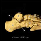
Talar Body Fracture S92.123A 825.21

Talus lateral view image 
ICD-9 Classification / Treatment
Etiology / Natural History Associated Injuries / Differential Diagnosis
Anatomy Complications
Clinical Evaluation Follow-up Care
Xray / Diagnositc Tests Review References

synonyms: Astragalus fracture, talar body fracture, talar fracture,

Talus Fracture ICD-10

A- initial encounter for closed fracture

B- initial encounter for open fracture

D- subsequent encounter for fracture with routine healing

G- subsequent encounter for fracture with delayed healing

K- subsequent encounter for fracture with nonunion

P- subsequent encounter for fracture with malunion

S- sequela

Talus Fracture ICD-9

  • 825.21(closed)
  • 825.31(open)

Talar Body Fracture Etiology / Epidemiology / Natural History

  • Uncommon
  • @20% of talus factures.
  • Talar body fractures = intraarticular fractures involving the tibiotalar and subtalar joint. Talar body fractures = inferior fracture line extends into or posterior to the lateral process. Talar neck fractures = fractures in which the inferior fracture line passes anterior to the lateral process of the talus in the region of the tarsal sinus. (Inokuchi S, Foot Ankle Int, 1996;17: 748).

Talar Body Fracture Anatomy

  • Blood supply: the artery of the tarsal canal(posterior tibial artery) is the main blood supply to the body of the talus.  It forms an anastomotic sling with the artery of the tarsal sinus (peroneal).  Others=superior neck vessels from anterior tibial artery, Deltoid artery(post tibial) (Mulfinger JBJS 1970;52B:160), (Gelberman RH, Foot Ankle 1983;4:64).
  • 60% covered with articular cartilage, no muscle originate from or insert into talus.
  • Lateral process: a wide, wedge-shaped prominence extending from the lateral aspect of the body of the talus; consists of two distinct articular facets: the dorsolateral and the inferomedial. The dorsolateral facet articulates with the distal fibula; the inferomedial facet forms the anterolateral portion of the subtalar joint. The lateral process is the site of insertion of the lateral talocalcaneal ligament.
  • Posterior process: composed of a medial and a lateral tubercle(Stieda’s process) which are separated by a groove within which lies the flexor hallucis longus tendon. The Y-shaped, bifurcate talocalcaneal ligament forms a roof over this grooveand inserts onto each tubercle. The posterior talofibular ligament inserts onto the lateral tubercle of the talus. The posterior talotibial portion of the deltoid ligament inserts onto the medial tubercle.
  • Os trigonum: located directly posterior to the lateral tubercle(Stieda's process). It is an accessory bone that arises from a secondary ossification center between the ages of 8 and 11 years. Generally fuses to the lateral tubercle within 1 year of its appearance. May persist as a separate ossicle, attached to the talus by a cartilaginous synchondrosis. (Grogan DP, JPO 1990;10:618)

Talar Body Fracture Clinical Evaluation

  • Ankle pain and swelling, inability to ambulate.
  • Evaluate soft tissue injury and for open fractures.
  • Document neurovascular exam before and after any treatment.

Talar Body Fracture Xray

  • A/P, Lateral, and Mortise Ankle and A/P, Oblique, Lateral foot xrays indicated.
  • Canale view-visualizes talar neck; taken with ankle in maximum plantar flexion and foot pronated 15 degrees. Radiograph directed at 75 degree angle from horizontal plane in the A/P plane. (Canale, JBJS 60A:143;1978).
  • Hawkins Sign=prognostic indicator following displaced neck fx’s. Subchondral radiolucency presnt 6-8 wks after fx suggests blood supply to this region.
  • MRI helpful to determing AVN post-operatively, but may be limited by metal from ORIF

Talar Body Fracture Classification / Treatment

  • ATLS resuscitation. These can be high enegery injuries, assessment should begin with the A,B,C's.
  • Isolated, Nondisplaced (<2mm displacement)
    -Treatment = NWB SLC 6-10 wks with frequent follow xrays to confirm maintenance of reduction. Consider internal fixation to allow early ROM and prevent loss of reduction.
  • Displaced (>2mm displacement ), or with associated injury (talar neck fracture):
    -high risk of AVN
    -Treatment = ORIF via medial of lateral approaches depending on fracture pattern. Fixation with small-fragment, mini-fragment stainless steel implants or headless compression screws. Consider medial malleolar osteotomy and limitied anterolateral arthrotomy.  Grob CORR 199:88;1985
  • Open fracture treatment depending on grade.

Talar Body Fracture Associated Injuries

  • Talar Neck fracture
  • Lateral process (snowboarder's fx)
  • Fracture-dislocation of the subtalar joint
  • Fracture-dislocation of the ankle
  • Ankle fracture
  • Metatarsal fracture
  • Ankle Sprain

Talar Body Fracture Complications

  • Osteonecrosis
  • Malunion
  • Nonuion
  • Infection
  • Painful hardware
  • CRPS
  • DVT / PE

Talar Body Fracture Follow-Up care

  • Nonweigthbearing cast in a plantigrade postion for 6-8 wks; followed by ROM exercises.
  • Nonweighbearing should continue for at least 3 months
  • 38% Osteonecrosis, 65% tibiotalar arthritis, 35% subtalar arthritis afetr operative treatment. (Vallier HA, JBJS 2003;85A:1716).

Talar Body Fracture Review References

  • Rockwood and Greens
  • Grob D, Simpson LA, Weber BG, Bray T: Operative treatment of displaced talus fractures.  Clin Orthop 1985;199:88-96
  • Vallier HA, JBJS 2003;85A:1716.

cc